3D model for cartilage degradation
Effect of mechanical stress on cartilage degradation studied in vitro
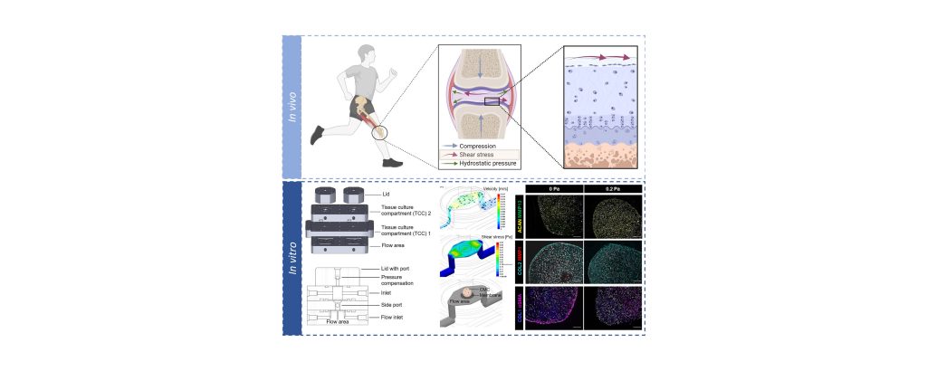
Dr. Alexandra Damerau and Dr. Timo Gaber (DRFZ and Charité – Universitätsmedizin Berlin) have used an innovative in-vitro 3D model to investigate the mechanical influences on cartilage tissue. They were able to show that mechanical stress can attenuate inflammation-induced cartilage degradation in osteoarthritis. They also demonstrated the anti-inflammatory effect of JAK inhibitors. Their model offers new approaches for the development of animal-free therapies. The results were published in the journal Advanced Science.
Researchers from the group of Prof. Dr. Frank Buttgereit (Charité – Universitätsmedizin Berlin), together with colleagues from Sanofi-Aventis Deutschland GmbH and the School of Life Sciences in Switzerland, developed a state-of-the-art in-vitro 3D model that mimics the natural environment of cartilage. This enabled them to precisely reproduce the complex mechanical forces that act on the cartilage in the joints. Using human chondrogenic microconstructs, the team simulated both healthy and pathological cartilage conditions and provided new insights into the role of mechanical stimuli on cartilage integrity. The study shows that mechanical stress can inhibit inflammation and thus counteract cartilage degradation.
The researchers also used this model to test the therapeutic potential of a JAK inhibitor to alleviate osteoarthritis-related damage. A key driver of joint inflammation in osteoarthritis is the cytokine TNF-α, the persistent activity of which ultimately leads to cartilage degradation. The anti-inflammatory effect of JAK inhibitors significantly reduced this degradation.
This innovative model not only provides crucial insights into the molecular mechanisms of osteoarthritis, but also paves the way for ethically acceptable, animal-free test procedures for novel therapies.