
Diefenbach lab
Development and function of the innate immune system
Developmental and Mucosal Immunology
Our current and future research is focused on a molecular understanding of how components of the innate immune system promotes tissue homeostasis by contributing to the adaptation of multicellular organisms to pernicious environments, such as those at barrier surfaces (e.g., intestine, skin).
We are addressing three major questions:
1. Transcriptional control of ILC development and function
Our lab has recently contributed to the discovery of innate lymphoid cells (ILCs), a group of tissue-resident innate lymphocytes located at border surfaces that release cytokines specifically acting on epithelial cells thereby contributing to tissue homeostasis and to the adaptation of the host in response to noxious compounds and tissue damage. A focus has been the analysis of transcriptional programs controlling lineage specification, commitment and function of ILCs. We recently identified a common, Id2-expressing progenitor to all ‘helper-like’ ILC lineages, the CHILP (Klose, Cell 2014). Our previous work has revealed that functional perturbations of ILC predispose to intestinal infections and to chronic inflammatory bowel diseases. It is likely, that such studies will identify new targets for the treatment of debilitating chronic inflammatory disorders.
2. The role of the indigenous microbiota in calibrating innate immunity
The role of environmental factors (microbiota, nutrients) for the formation of an effective and well calibrated innate immune response is recognized. Mononuclear phagocytes germ-free mice were unable to produce most pro-inflammatory cytokines (in particular type I IFN), and, consequently, germ-free mice were more susceptible to infections with viruses (Ganal, Immunity 2012). On a mechanistic level, we showed that signals of the microbiota are needed to remove a chromatin barrier required for the transcription of genes after ligation of pattern recognition receptors (PRR). Ongoing studies address the mechanistic underpinnings of this process as well as the microbiota-derived signals that tune mononuclear phagocyte responses. As many autoimmune diseases have a type I IFN signature, it is possible that dyscalibration of the mononuclear phagocytes by the microbiota may contribute to rheumatic diseases.
3. The role of nutrients for development and function of the innate immune system
Much has been learned about how the microbiota contributes to many aspects of host physiology. In contrast, the role of nutrients for development and function of the intestinal immune system has largely been a matter of speculation owing to the fact that molecular sensors of dietary molecules are widely unknown. Given the broad role of nutrients in metabolic diseases and the impact of intestinal cancer on human health, research into the question of how the power of nutrients can be harnessed for improving human health and for the prevention of disease is much warranted. We have recently found that the aryl hydrocarbon receptor (AhR), a transcription factor activated by small molecular ligands, is required for the development of innate immune system components by serving as a sensor for phytochemicals. Based on these preliminary data, we aim now to systematically define the role of diet-induced changes for the function and differentiation of mucosa-associated innate lymphocytes and to uncover how innate lymphocytes regulate epithelial adaptation by controlling niche support for intestinal epithelial stem cells.
Microbiome
Innate immune system
Chronic inflammation
Tissue homeostasis
Innate Lymphoid Cells, ILC
Link to website at Charité: http://charite-mikrobiologie.de/diefenbach-lab/

Group leader
Univ. Prof. Dr. med. Andreas Diefenbach
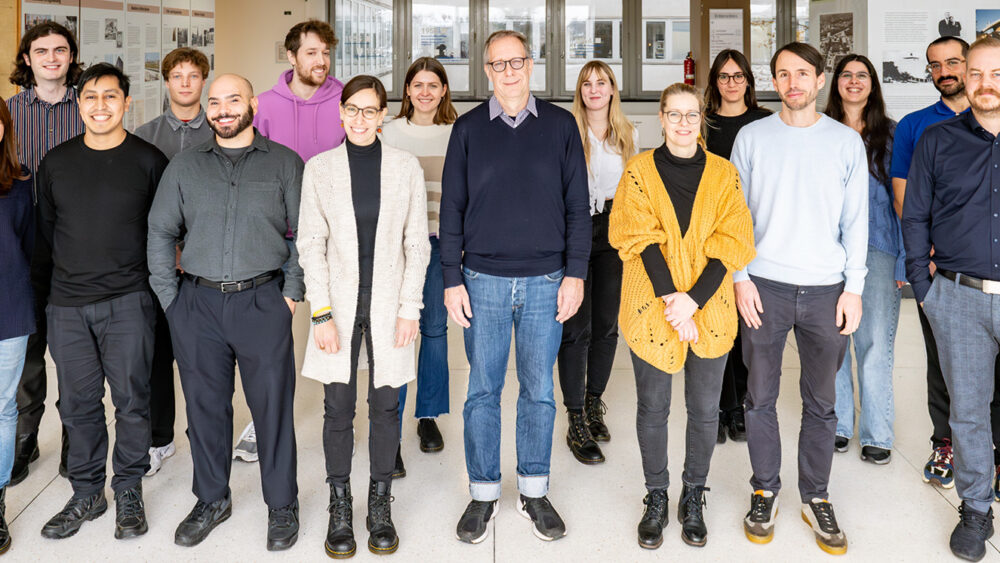
Group Members
Anna Fagundes
Stylianos Gnafakis
Luke Houghton
Atsuyo Ikeda
Luise Kausch-Blecken
Michael Kofoed-Branzk
Anita Kowalczyk
Irene Mattiola
Diego Perez Vazquez
Almina Polinova
Bruno Rocha
Omer Shomrat
Caroline Tizian
Nilsu Turay
Mario Witkowski
Original Articles
Witkowski, M., C.Tizian, M.Ferreira-Gomes, D.Niemeyer, T.C.Jones, F.Heinrich, S.Frischbutter, S.Angermair, T.Hohnstein, I.Mattiola, P.Nawrath, S.Mc Ewen, S.Zocche, E.Viviano, G.A.Heinz, M.Maurer, U.Kölsch, R.L.Chua, T.Aschman, C.Meisel, J.Radke, B.Sawitzki, J.Roehmel, K.Allers, V.Moos, T.Schneider, L.Hanitsch, M.A.Mall, C.Conrad, H.Radbruch, C.U.Duerr, J.A.Trapani, E.Marcenaro, T.Kallinich, V.M.Corman, F.Kurth, L.E.Sander, Christian Drosten, S.Treskatsch, P.Durek, A.Kruglov, A.Radbruch, M.F.Mashreghi, and A.Diefenbach. 2021. Untimely TGFb responses in severe COVID-19 limit antiviral function of NK cells. Nature. 600:295-301
Guendel, F., M.Kofoed-Branzk, K.Gronke, C.Tizian, M.Witkowski, H-W.Cheng, G.A.Heinz, F.Heinrich, P.Durek, P.S.Norris, C.F.Ware, C.Ruedl, S.Herold, K.Pfeffer, T.Hehlgans, A.Waisman, B.Becher, A.D.Giannou S.Brachs, K.Ebert, Y.Tanriver, B.Ludewig, M-F.Mashreghi, A.A.Kruglov, and A.Diefenbach. 2020. Group 3 innate lymphoid cells program a distinct subset of IL-22BP-producing dendritic cells demarcating solitary intestinal lymphoid tissues. Immunity. 53:1015-1032.
Schaupp L, S.Muth, L.Rogell, M.Kofoed-Branzk, F.Melchior, S.Lienenklaus, S.C.Ganal-Vonarburg, M.Klein, F.Guendel, T.Hain, K.Schütze, U.Grundmann, V.Schmitt, M.Dorsch, J.Spanier, P.Tegtmeyer, T.Schwanz, S.Jäckel, C.Reinhardt, T.Bopp, S.Danckwardt, K.Mahnke, G.A.Heinz, M.F.Mashreghi, P.Durek, U.Kalinke, O.Kretz, T.B.Huber, S.Weiss, C.Wilhelm, A.J.Macpherson, H.Schild, A.Diefenbach1,*, H.C.Probst*. 2020. Microbiota-induced tonic type I interferons instruct a poised basal state of dendritic cells. Cell. 181:1080-1096. 1Lead Senior Author; *equal contribution.
Gronke, K., P.P.Hernández, J.Zimmermann, C.S.N.Klose, M.Kofoed-Branzk, F.Guendel, M.Witkowski, C.Tizian, L. Amann, F.Schumacher, H.Glatt, A.Triantafyllopoulou, and A.Diefenbach. 2019. Interleukin-22 protects intestinal stem cells against genotoxic stress. Nature. 566:249-253.
Herrtwich, L., I.Nanda, K.Evangelou, T.Nikolova, V.Horn, Sagar, D.Erny, J.Stefanowski, L.Rogell, C.Klein, K.Gharun, M.Follo, B.Kremer, M.Seidl, N.Münke, J.Sanger, M.Fliegauf, T.Aschman, D.Pfeifer, S.Sarrazin, M.Sieweke, D.Wagner, C.Dierks, T.Haaf, T.Ness, M.M.Zaiss, R.E.Voll, S.D.Deshmukh, M.Prinz, T.Goldmann, C.Hölscher, A.E.Hauser, A.J.Lopez-Contreras, D.Grün, V.Gorgoulis, A.Diefenbach*, P.Henneke, and A.Triantafyllopoulou*. 2016. DNA damage signaling instructs polyploid macrophage fate in granulomas. Cell. 167:1264-1280. *co-corresponding authors
Review Articles
Diefenbach, A., S.Gnafakis, and O.Shomrat. 2020. Innate lymphoid cell-epithelial cell modules sustain intestinal homeostasis. Immunity. 52:452-463.
Mattiola, I., and A.Diefenbach. 2019. Innate Lymphoid Cells and Cancer at Border Surfaces With the Environment. Semin Immunol. 41:101278.
Branzk, N., K.Gronke, and A.Diefenbach. 2018. Innate lymphoid cells, mediators of tissue homeostasis, adaptation and disease tolerance. Immunol Rev. 286:86-101.
Vivier, E., D.Artis, M.Colonna, A.Diefenbach, J.P.Di Santo, G.Eberl, S.Koyasu, R.M.Locksley, A.N.McKenzie, R.E.Mebius, F.Powrie, and H.Spits. 2018. Innate lymphoid cells: ten years on. Cell. 174:1054-1066. (all authors contributed equally).
Hernandez, P., K.Gronke, and A.Diefenbach. 2018. A catch-22: Interleukin-22 and cancer. Eur J Immunol. 48:15-31.
Diefenbach, A., M.Colonna, and S.Koyasu. 2014. Development, differentiation and diversity of innate lymphoid cells. Immunity. 41:354-365.
Spits, H., D.Artis, M.Colonna, A.Diefenbach, J.P.Di Santo, G.Eberl, S.Koyasu, R.M.Locksley, A.N.McKenzie, R.E.Mebius, F.Powrie, and E.Vivier. 2013. Innate lymphoid cells – a proposal for uniform nomenclature. Nat. Rev. Immunol. 13:145-149. (all authors contributed equally).

 Deutsch