
Niesner lab
We develop technologies for intravital imaging and live cell imaging to achieve new insights into chronic inflammatory processes
Biophysical Analytics
We are developing multi-photon microscopy and multi-modal optical imaging methodology to investigate the cellular mechanisms of rheumatic and chronic inflammatory pathologies in living mice and tissues. We aim to provide quantification tools of not only cellular phenotype in organ context, in a time-lapse manner, but also of cellular and tissue function and dysfunction at highest optical performance in vivo, in mouse disease models. In this sense, fluorescence lifetime imaging of cells and, intravitally, of tissues allowed us to establish the concept of oxidative stress memory during the course of neuroinflammation, to identify factors maintaining it and, consequently, possible therapeutic approaches even in late stages of the disease – in close collaboration with our clinical partners.
This project is a collaboration with Dr. H. Radbruch and Prof. Dr. F. Heppner (Neuropathology, Charité), Prof. Dr. F. Paul (Neuroimmunology, Charité) and Prof. Dr. A.E. Hauser (DRFZ). Since we expect a similar phenotypic shift in renal tissue of mice affected by lupus, we are currently performing longitudinal NAD(P)H fluorescence lifetime imaging of the kidney in collaboration with Prof. Dr. R. Voll (Erlangen, CRC130). We expanded our technology also to other organs and cell types such as the bone marrow.
Besides, taking into account the fact that the time course of chronic inflammatory diseases as well as the underlying dynamic mechanisms are not fully understood, we develop longitudinal imaging technologies to comprehend these mechanisms within one and the same individual, at cellular level.
To establish the link between the thorough knowledge we gain from our dynamic and functional intravital microscopic investigations and parameters measurable in a clinical setting, we combine non-invasive, label-free imaging methods available in clinics (such as the optical coherence tomography) with our multi-photon imaging approaches.
We aim to apply these tools in the context of rheumatic disease models to better correlate pathological mechanisms revealed by intravital microscopy with typical parameters of tissue degeneration revealed by clinical diagnosis.
Keywords
Intravital multi-photon microscopy
NAD(P)H metabolism
Functional in vivo imaging

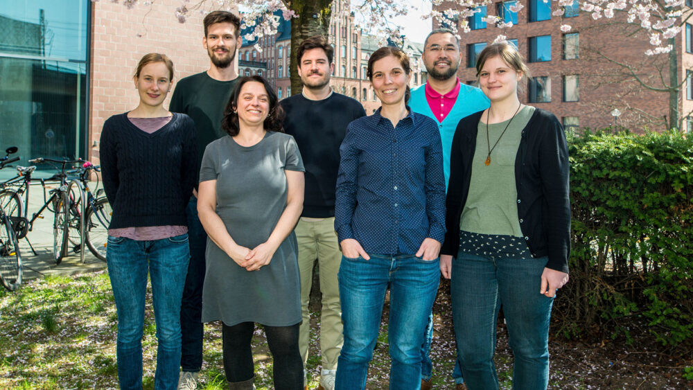
Group leader
Prof. Dr. rer. nat. Raluca A. Niesner
Scientists
Dr. Ruth Leben
PhD students
Alexander Fiedler
Anne Bias
Danja Brandt
Technician
Robert Günther
Volker Andresen, LaVision Biotec – Milteny, Bielefeld/Germany
Dr. Marcus Beutler, APE, Berlin/Germany
Prof. Georg Duda, Julius-Wolf-Institute, Charité /Berlin/Germany
Dr. David Entenberg, Albert Einstein College of Medicine/Intravital Imaging/ New York/US
Prof. Dr. Susanne Hartmann, Freie Universität, Berlin /Veterinary Medicine/Berlin/Germany
Prof. Dr. Anja E. Hauser, DRFZ and Charité/Immune Dynamics/ Berlin/Germany
Prof. Dr. Frank Heppner, Charité /Neuropathology/Berlin/Germany
Dr. Julia Jellusowa, University Freiburg/Faculty for Biology/Freiburg/Germany
Romano Matthys, RISystem, Davos/Switzerland
Prof. Dr. Dirk Mielenz, University Nürenberg-Erlangen / Immunology/ Erlangen/Germany
Prof. Dr. Friedemann Paul, Charité /Neuroimmunology/Berlin/Germany
Dr. Helena Radbruch, Charité /Neuropathology/Berlin/Germany
Prof. Dr. Tim Schulz, DIfE / Potsdam/Germany
Dr. Heinrich Spiecker, LaVision Biotec – Milteny, Bielefeld/Germany
Prof. Dr. Reinhard Voll, University Hospital Freiburg/Rheumatology/Freiburg/Germany
Prof. Dr. Carolina Wählby, Uppsala University/Quantitative Microscopy/Upsalla/Sweden
Prof. Dr. Jürgen Zentek, Freie Universität, Berlin / Veterinary Medicine /Berlin/Germany
DFG: TRR130, C01
DFG: FOR2165-2, TP7
Einstein Foundation: Collaborative project with Prof. A.E. Hauser and Prof. M. Leisegang, on T-cell therapies in Multi-Myeloma (Charité).
- “Teriflunomide does not change dynamics of NADPH oxidase activation and neuronal dysfunction during neuroinflammation.” Mothes, C. Ulbricht, R. Leben, R. Günther, A.E. Hauser, H. Radbruch*, R.A. Niesner*. Front. Mol. Biosci. 2020.
- “B cell speed and B-FDC contacts in germinal centers determine plasma cell output via Swiprosin-1/EFhd2.” Reimer D., Meyer-Hermann M., Rakhymzhan A., Steinmetz T., Tripal P., Thomas J., Boettcher M., Mougiakakos D., Schulz S.R., Urbanczyk S., Hauser A.E., Niesner R.A.*, Mielenz D.* Cell Reports.
- “Co-registered spectral optical coherence tomography and two-photon microscopy for multimodal near-instantaneous deep-tissue imaging.” Rakhymzhan, L. Reuter, R. Raspe, D. Bremer, R. Günther, R. Leben, J. Heidelin, V. Andresen, S. Cheremukhin, H. Schulz-Hildebrandt, M.G. Bixel, R.H. Adams, H. Radbruch, G. Hüttmann, A.E. Hauser, R.A. Niesner. Cytometry Part A.2020 May.97(5):515-527.doi: 10.1002/cyto.a.24012.
- „Systematic Enzyme Mapping of Cellular Metabolism by Phasor-Analyzed Label-Free NAD(P)H Fluorescence Lifetime Imaging.“ Leben*, M. Köhler*, H. Radbruch, A.E. Hauser, R. Niesner. IJMS. 2019.
- “Synergistic Strategy for Multicolor Two-photon Microscopy: Application to the Analysis of Germinal Center Reactions In Vivo.“ Rakhymzhan A, Leben R, Zimmermann H, Günther R, Mex P, Reismann D, Ulbricht C, Acs A, Brandt AU, Lindquist RL, Winkler TH, Hauser AE, Niesner RA. Sci Rep. 2017 Aug 2;7(1):7101. doi: 10.1038/s41598-017-07165-0.
- “Longitudinal intravital imaging of the femoral bone marrow reveals plasticity within marrow vasculature.“ Reismann D, Stefanowski J, Günther R, Rakhymzhan A, Matthys R, Nützi R, Zehentmeier S, Schmidt-Bleek K, Petkau G, Chang HD, Naundorf S, Winter Y, Melchers F, Duda G, Hauser AE*, Niesner RA*. Nat Commun. 2017 Dec 18;8(1):2153. doi: 10.1038/s41467-017-01538-9.
- „Phasor-Based Endogenous NAD(P)H Fluorescence Lifetime Imaging Unravels Specific Enzymatic Activity of Neutrophil Granulocytes Preceding NETosis.“ Leben R, Ostendorf L, van Koppen S, Rakhymzhan A, Hauser AE, Radbruch H, Niesner RA. Int J Mol Sci. 2018 Mar 29;19(4). pii: E1018. doi: 10.3390/ijms19041018.

 Deutsch
